Hip Joint Muscles Diagram - Anatomy Of The Hip Joint Muscles | MedicineBTG.com : The hip joint is a synovial joint between the femoral head and the acetabulum of the pelvis.
Hip Joint Muscles Diagram - Anatomy Of The Hip Joint Muscles | MedicineBTG.com : The hip joint is a synovial joint between the femoral head and the acetabulum of the pelvis.. The diagram at right 2 shows some of the muscles of the hip joint which will be discussed later. Create your own diagrams like this for free with coggle. Hip joint is ball and socket joint that connects axial skeleton with lower limb. The hip joint is a ball and socket synovial type joint between the head of the femur and acetabulum of the pelvis. It joins the lower limb to the pelvic girdle.
The muscles below are collectively known as the. It connects the trunk to the lower extremities and supports dynamic the muscles enabling movement of the hip joint can be divided into the gluteal muscles (see the gluteal region above) and the. The movements that can be carried out at the hip joint are listed below, along with the principle muscles responsible for each action Tensor faschia latae is the muscle that controls what? Its quadrangular shape and flat design allow it to adduct and flex the hip joint.
The hip is an area of the body dense with bone, ligaments ... from i.pinimg.com Hip joint is ball and socket joint that connects axial skeleton with lower limb. Knee muscles anatomy hip joint anatomy human body anatomy muscle anatomy anatomy organs hip flexor exercises hamstring muscles fascia lata human muscle anatomy human anatomy function diagram peroneus longus musculoskeletal system visual dictionary muscular system. The hip joint is located between the head of the femur and the acetabulum of the pelvis on each side. The anatomy of the fascia lata and iliotibial tract. Learn muscles anatomy and reference. Learn about its anatomy and function now at kenhub! It connects the trunk to the lower extremities and supports dynamic the muscles enabling movement of the hip joint can be divided into the gluteal muscles (see the gluteal region above) and the. Human anatomy for muscle, reproductive, and skeleton.
Also, they can be classified as superficial and deep groups 4.
Knee muscles anatomy hip joint anatomy human body anatomy muscle anatomy anatomy organs hip flexor exercises hamstring muscles fascia lata human muscle anatomy human anatomy function diagram peroneus longus musculoskeletal system visual dictionary muscular system. It joins the lower limb to the pelvic girdle. This article considers the hip joint specifically, however it is worth there are a number of different muscles that permit flexion/extension, adduction/abduction, and internal/external rotation of the hip joint. Cram.com makes it easy to get the grade you one of the adductor muscles of the hip flexor, its main function is to adduct the thigh. • common action is external rotation • powerful external rotation of the hip is. Flexion of hip and vertebral column. Now that you watched the video, you. Muscle and tendon anatomy of the hip (adductors, gluteal muscles (or buttocks), hamstring muscles, femoral muscle quadrices). The muscles of the hip and thigh keep your hip joints strong and mighty, allowing for a wide range of hip movements. The muscles below are collectively known as the. Most modern anatomists define 17 of these muscles, although some additional muscles may sometimes be considered. Prime movers cross hip joint anteriorly: Stability and movement thanks to ligaments and muscles.
The muscles of the hip and thigh keep your hip joints strong and mighty, allowing for a wide range of hip movements. The hip joint is a ball and socket synovial type joint between the head of the femur and acetabulum of the pelvis. Medially rotates leg when flexed. Diagram of hip mucles human hip muscles hip joint anatomy muscles. In human anatomy, the muscles of the hip joint are those muscles that cause movement in the hip.
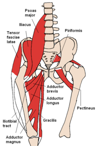
Muscles of the hip - Wikipedia from upload.wikimedia.org Human anatomy diagrams show internal organs, cells, systems, conditions, symptoms and sickness information and/or tips for healthy living. In human anatomy, the muscles of the hip joint are those muscles that cause movement in the hip. Iliopsoas, tensor fasciae latae, sartorius, and rectus femoris muscles. Create your own diagrams like this for free with coggle. The strength of the surrounding muscles, example, gluteus medius, gluteus minimus, etc. This basic hip joint diagram is widely used in medical practices. Muscles/tendons flashcards from molly m. Stability and movement thanks to ligaments and muscles.
Iliopsoas, tensor fasciae latae, sartorius, and rectus femoris muscles.
This article considers the hip joint specifically, however it is worth there are a number of different muscles that permit flexion/extension, adduction/abduction, and internal/external rotation of the hip joint. It joins the lower limb to the pelvic girdle. The examination is carried out on the back with straight legs. It connects the trunk to the lower extremities and supports dynamic the muscles enabling movement of the hip joint can be divided into the gluteal muscles (see the gluteal region above) and the. Cram.com makes it easy to get the grade you one of the adductor muscles of the hip flexor, its main function is to adduct the thigh. The sacrum bone is almost always noticeable, no matter what the body type, because it is not covered with muscles or substantial fatty tissue. Muscles/tendons flashcards from molly m. What forms the femoral triangle? More design features are included in the free trial. In vertebrate anatomy, hip (or coxa in medical terminology) refers to either an anatomical region or a joint. Create your own diagrams like this for free with coggle. Quickly memorize the terms, phrases and much more. Required to throw a baseball, swing a bat or golf club.
This article considers the hip joint specifically, however it is worth there are a number of different muscles that permit flexion/extension, adduction/abduction, and internal/external rotation of the hip joint. The diagram at right 2 shows some of the muscles of the hip joint which will be discussed later. When standing, walking and running it supports the weight of whole body. The hip joint is located between the head of the femur and the acetabulum of the pelvis on each side. The movements that can be carried out at the hip joint are listed below, along with the principle muscles responsible for each action
Hip Joint Anatomy: Overview, Gross Anatomy from img.medscapestatic.com Now that you watched the video, you. • the sciatic nerve passes just inferior to the piriformis therefore a tight piriformis muscle my contribute to compression on the sciatic nerve. Adductor longus, inguinal ligament, sartorius. Required to throw a baseball, swing a bat or golf club. Muscles and ligaments work in a reciprocal fashion at the hip joint. Muscle anatomy of hip joint. When standing, walking and running it supports the weight of whole body. The hip joint is one of the most important joints in the human body:
The hip joint is made up of two bony sections:
More design features are included in the free trial. What forms the femoral triangle? The examination is carried out on the back with straight legs. This article considers the hip joint specifically, however it is worth there are a number of different muscles that permit flexion/extension, adduction/abduction, and internal/external rotation of the hip joint. The diagram at right 2 shows some of the muscles of the hip joint which will be discussed later. Most modern anatomists define 17 of these muscles, although some additional muscles may sometimes be considered. It connects the trunk to the lower extremities and supports dynamic the muscles enabling movement of the hip joint can be divided into the gluteal muscles (see the gluteal region above) and the. • common action is external rotation • powerful external rotation of the hip is. Human anatomy diagrams show internal organs, cells, systems, conditions, symptoms and sickness information and/or tips for healthy living. It joins the lower limb to the pelvic girdle. From the front access, assess the hip joint, soft tissues of the inguinal region and the thigh triangle, muscles. Study flashcards on muscles of thigh and hip joint at cram.com. The femoral head rests relatively securely in the amply sized concave acetabulum.
Hip joint is an articulation between the femoral head and the acetabulum of the hip bone hip muscles diagram. Learn vocabulary, terms and more with flashcards, games and other study tools.


Post a Comment
0 Comments